オトガイ形成(Genioplasty)

◆オトガイ形成(Genioplasty)◆
オトガイは正面顔の輪郭における最下端に位置しており、顔が完成されるエンドポイントになります。このため、オトガイは顔面の輪郭形成術において最も古くから注目されており、様々な報告がなされています。また、オトガイの非対称や変形は容貌とそれにともなう心の変化に強い影響を持つとされています1)。オトガイ伸びて長いと顔の重心が下に偏り、顔が大きく、鈍い印象を与えます。一方で、オトガイが小さすぎる場合にも弱々しくイメージになりがちです。オトガイは顔全体の調和を取るのに非常に重要な役割を果たしています2-5)。オトガイの多様多彩な形態に対し骨切り術を行い、個々人に最も似合うオトガイを形成することで顔の全体的な印象を改善させることができます5)6)。また、前述のようにオトガイは顔面のエンドポイントであることから、顎矯正手術や他の輪郭形成術と組み合わせて行うとより効果的な審美的改善効果が得られるとされています7-10)。
◆世界におけるオトガイ形成術の動向◆
日本を含むアジア圏では四角くて、大きく丸い顔は魅力的ではないとされています。そのため特に若い女性は卵型でアーモンド形の顔や、シャープな顎を好む傾向にあります11)。これを受けて日本、韓国、台湾ではオトガイ形成術は美容外科手術の中でも最もよく行われる手術の一つになっています。
◆様々なオトガイ形成術◆
一言にオトガイ形成術といっても、目的によりさまざまな手法や骨切りの方法があります。
オトガイの前方移動や後方移動12)、高さや長さの調節13)14)、幅の調節15)16)などです。水平骨切り術(Horizontal osteotomy)13)17)18)、Wedge osteotomy19)、Curving osteotomy20)、M字型オトガイ形成21)、矢状分オトガイ形成22)、幅径縮小分節オトガイ形成23)、Basilar骨切り術24)、ZigZag骨切り術25)など様々なバリエーションが報告されています。また、その治療目的ですが、整容的な改善のみならず、口唇機能の最適化26)、睡眠時無呼吸27-29)の治療の一環として行われることもあります。
◆治療のポイントと下顎の輪郭形成の種類◆
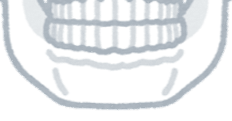
・下顎はオトガイ部、体部、角部、下顎枝、関節突起、筋突起より構成されます。
・下顎の輪郭形成はこれらの各部位を切除したり、削ったり、移動して再固定することにより行います。
◆オトガイ形成をおすすめの方◆
・オトガイの形に不満足なすべての方(非対称、大きすぎ、小さすぎなど)
・オトガイの後退がある方
・オトガイが長い方
・オトガイが短い方
◆オトガイ形成術の種類と手術部位◆
・オトガイ形成術には下記のような種類があります。
・複数の種類のオトガイ形成に加えて、下顎形成術を併用してより効果的な治療が行われます。
①オトガイ前方移動
・小さくて短いオトガイ(オトガイがない方)を前方に移動させて、横顔がきれいになるようなオトガイを形成します。
②オトガイ中抜き
・長いオトガイを短くします。
➂オトガイ延長
・小さくて短いオトガイ(オトガイがない方)を前下方向に移動させて、横顔がきれいになるようなオトガイを形成します。骨の移植が必要になることがあります。
◆手術◆
①口腔内の切開
・口腔内を切開して目的の手術部位(オトガイ部)へ到達します。
②骨切りラインのデザイン
・術前に行った術前シミュレーションにを基にして、骨切りラインの設定を行います。
➂骨切り
・顔面骨骨切り用の専用の機械で、目的部位の骨切りを行います。
④骨の移動とプレート固定
・骨を予定の位置に移動させて、プレートで固定します。
⑤縫合
・創部を縫合します。
◆「オトガイ形成」に特異的なダウンタイム、術後経過、合併症◆
●オトガイ神経麻痺(しびれ)30)31)
・下顎の骨切りの際に、オトガイ神経の麻痺が生じる可能性があります。オトガイ神経は肉眼で確認可能ですが、非常に弱い神経であるため麻痺が出やすい神経です。
・神経を切断することはほとんどありませんが、骨切り線を確保する際に剥離操作を行ったり、筋鈎で引いたするすることがあります。
・オトガイ部~下口唇、下の歯茎の知覚鈍麻や知覚脱失が生じます。
●顔面神経頬骨枝及び下顎縁枝麻痺
・口の周りを動かす筋肉を支配している神経です。
・骨から筋肉を剥離する際に、損傷する可能性があります。
・十分に気を付けて行うため、切断のリスクは高くありませんが、骨切り線を確保する際に剥離操作を行ったり、筋鈎で引いたする際に圧迫されることがあります。
・口を動かす操作がしばらく困難になります。
●骨の移動量と軟部組織の変化●
・骨の移動量と軟部組織の移動量は一致しません32)。骨の移動量に対する軟部組織の変化については様々なシミュレーションのもとに検証が行われていますが、いまだ一定の見解を得ていないのが現状です2)5)17)33)。一般的には水平方向の変化は骨の移動量が1の場合、軟部組織の変化は0.7-0.9を基準としてプランニングを行います34)35)。しかし、前述のように十分にシミュレーションができないため、事前のプランニングに加えて、手術中にフェイスラインの変化具合をみながらの判断が重要となります。
●下唇の形態変化と軟部組織の変化●
・オトガイ形成により下唇の周囲の筋肉の移動と再付着が生じます。
・下唇が薄くなったり、厚くなったりする変化が起きることがあります。
・オトガイ唇溝が強調されたり、オトガイ筋の過度な収縮によりオトガイの軟部組織の形態が変化することがあります。
●強調されたオトガイ唇溝の修正
・シリコンプロテアーゼの挿入などの処置があります。
●オトガイの下垂(chin ptosis)とwitch’s chin●
・オトガイ部分が過度に下垂したり、魔女の顎のように異常に突出して段差のついた顎になることがあります。これは、オトガイ部分の筋肉の過度な剥離や予定しない部分への再付着が原因とされています。手術操作の際にできるだけ剥離を少なくすることで予防可能とされています32)。しかし、変形したオトガイの処理が必要な場合、骨の中抜きが必要な場合、顔面女性化手術の手術などでは、骨の処理のために広範な剥離が必要となります32)。その場合には、筋肉の再固定を行うなど工夫を行うことによりオトガイ部分の下垂の予防を行います36-39)。
●オトガイ形成とシリコンインプラント●
・オトガイ部分へのシリコンインプラントは前方向へのオトガイの調節しか行うことができませんが、オトガイ形成術ではオトガイ部分を6方向(垂直面、矢状面、水平面)に移動可能です17)40)。
●オトガイ形成術と骨移植●
・オトガイ延長の場合には骨移植が必要となります。骨は頭蓋骨や腸骨から採取します。同時に顔面の他の部位の手術(下顎短縮など)を行う場合はその部分から骨を採取することもあります。
●オトガイ形成術と睡眠時無呼吸●
・睡眠時無呼吸(OSAS:obstructive sleep apnea syndrome)
・オトガイの前方移動の場合は、後退した顎の筋肉を前方に牽引することが可能であるため、オトガイ後退の治療と同時に睡眠時無呼吸の治療効果があります27)41)。
◆参考文献◆
1) Spear S. L., Kassan M.: Genioplasty. Clin Plast Surg 16:695-706, 1989
2) San Miguel Moragas J., Oth O., Büttner M.et al: A systematic review on soft-to-hard tissue ratios in orthognathic surgery part II: Chin procedures. J Craniomaxillofac Surg 43:1530-1540, 2015
3) Abadi M., Pour O. B.: Genioplasty. Facial Plast Surg 31:513-522, 2015
4) Arcas A., Vendrell G., Cuesta F.et al: Advantages of performing mentoplasties with customized guides and plates generated with 3D planning and printing. Results from a series of 23 cases. J Craniomaxillofac Surg 46:2088-2095, 2018
5) Möhlhenrich S. C., Heussen N., Kamal M.et al: Influence of setback and advancement osseous genioplasty on facial outcome: A computer-simulated study. J Craniomaxillofac Surg 43:2017-2025, 2015
6) Gonzalez-Ulloa M.: Quantitative principles in cosmetic surgery of the face (profileplasty). Plast Reconstr Surg Transplant Bull 29:186-198, 1962
7) Chang E. W., Lam S. M., Karen M.et al: Sliding genioplasty for correction of chin abnormalities. Arch Facial Plast Surg 3:8-15, 2001
8) Hoenig J. F.: Sliding osteotomy genioplasty for facial aesthetic balance: 10 years of experience. Aesthetic Plast Surg 31:384-391, 2007
9) Schwitzer J. A., Albino F. P., Mathis R. K.et al: Assessing Patient-Reported Outcomes Following Orthognathic Surgery and Osseous Genioplasty. J Craniofac Surg 26:2293-2298, 2015
10) Park J. Y., Kim M. J., Hwang S. J.: Soft tissue profile changes after setback genioplasty in orthognathic surgery patients. J Craniomaxillofac Surg 41:657-664, 2013
11) Nocini P. F., Chiarini L., Bertossi D.: Cosmetic procedures in orthognathic surgery. J Oral Maxillofac Surg 69:716-723, 2011
12) Hinds E. C., Kent J. N.: Genioplasty: the versatility of horizontal osteotomy. J Oral Surg 27:690-700, 1969
13) Converse J. M., Wood-Smith D.: HORIZONTAL OSTEOTOMY OF THE MANDIBLE. Plast Reconstr Surg 34:464-471, 1964
14) Anquetil M., Perrin J. P., Praud M.et al: Vertical lengthening genioplasty: A new osteotomy technique. J Stomatol Oral Maxillofac Surg 121:159-162, 2020
15) Bell W. H., Gallagher D. M.: The versatility of genioplasty using a broad pedicle. J Oral Maxillofac Surg 41:763-769, 1983
16) Raffaini M., Sesenna E.: Hemi-genioplasty: a technique to correct chin asymmetry. J Oral Maxillofac Surg 53:1362-1364, 1995
17) Sarver D. M., Johnston M. W.: Orthognathic surgery and aesthetics: planning treatment to achieve functional and aesthetic goals. Br J Orthod 20:93-100, 1993
18) Jones B. M., Vesely M. J.: Osseous genioplasty in facial aesthetic surgery--a personal perspective reviewing 54 patients. J Plast Reconstr Aesthet Surg 59:1177-1187, 2006
19) Costa P. J. C., de Gauw J. H., Costa Filho J. Z.: Wedge Osteotomy for Correction of Chin Asymmetry. J Craniofac Surg 29:e190, 2018
20) Wang J., Gui L., Xu Q.et al: The sagittal curving osteotomy: a modified technique for advancement genioplasty. J Plast Reconstr Aesthet Surg 60:119-124, 2007
21) Fariña R., Valladares S., Aguilar L.et al: M-shaped genioplasty: a new surgical technique for sagittal and vertical chin augmentation: three case reports. J Oral Maxillofac Surg 70:1177-1182, 2012
22) Schendel S. A.: Sagittal split genioplasty: a new technique. J Oral Maxillofac Surg 68:931-934, 2010
23) Uckan S., Soydan S., Veziroglu F.et al: Transverse reduction genioplasty to reduce width of the chin: indications, technique, and results. J Oral Maxillofac Surg 68:1432-1437, 2010
24) Li X., Hsu Y., Hu J.et al: Comprehensive consideration and design for treatment of square face. J Oral Maxillofac Surg 71:1761.e1761-1714, 2013
25) Keyhan S. O., Khiabani K., Hemmat S.et al: Zigzag genioplasty: a new technique for 3-dimensional reduction genioplasty. Br J Oral Maxillofac Surg 51:e317-318, 2013
26) Triaca A., Furrer T., Minoretti R.: Chin shield osteotomy--a new genioplasty technique avoiding a deep mento-labial fold in order to increase the labial competence. Int J Oral Maxillofac Surg 38:1201-1205, 2009
27) Riley R. W., Powell N. B.: Maxillofacial surgery and obstructive sleep apnea syndrome. Otolaryngol Clin North Am 23:809-826, 1990
28) Riley R., Guilleminault C., Powell N.et al: Mandibular osteotomy and hyoid bone advancement for obstructive sleep apnea: a case report. Sleep 7:79-82, 1984
29) Mehra P., Wolford L. M.: Surgical management of obstructive sleep apnea. Proc (Bayl Univ Med Cent) 13:338-342, 2000
30) Gianni A. B., D'Orto O., Biglioli F.et al: Neurosensory alterations of the inferior alveolar and mental nerve after genioplasty alone or associated with sagittal osteotomy of the mandibular ramus. J Craniomaxillofac Surg 30:295-303, 2002
31) Westermark A., Bystedt H., von Konow L.: Inferior alveolar nerve function after mandibular osteotomies. Br J Oral Maxillofac Surg 36:425-428, 1998
32) Zhang B. H., Byrd R., Bradley C.et al: Osseous Genioplasty: Prevention of Witch's Chin Deformity with No-Degloving Technique. Plast Reconstr Surg 148:720e-726e, 2021
33) Bell R., Kiyak H. A., Joondeph D. R.et al: Perceptions of facial profile and their influence on the decision to undergo orthognathic surgery. Am J Orthod 88:323-332, 1985
34) Ewing M., Ross R. B.: Soft tissue response to mandibular advancement and genioplasty. Am J Orthod Dentofacial Orthop 101:550-555, 1992
35) Shaughnessy S., Mobarak K. A., Høgevold H. E.et al: Long-term skeletal and soft-tissue responses after advancement genioplasty. Am J Orthod Dentofacial Orthop 130:8-17, 2006
36) Morris D. E., Lo L. J., Margulis A.: Pitfalls in orthognathic surgery: avoidance and management of complications. Clin Plast Surg 34:e17-29, 2007
37) White J. B., Dufresne C. R.: Management and avoidance of complications in chin augmentation. Aesthet Surg J 31:634-642, 2011
38) Chan D., Ducic Y.: A Simplified, Reliable Approach for Advancement Genioplasty. JAMA Facial Plast Surg 18:114-118, 2016
39) Frodel J. L., Jr., Rudderman R.: Facial soft tissue resuspension following upper facial skeletal reconstruction. J Craniomaxillofac Trauma 2:24-30, 1996
40) HK. Kawamoto: Osseous genioplasty. Aesthet Surg J 20:509-516, 2000
41) Precious D. S., Delaire J.: Correction of anterior mandibular vertical excess: the functional genioplasty. Oral Surg Oral Med Oral Pathol 59:229-235, 1985
この記事が気に入ったらサポートをしてみませんか?