疑問視されるSV40癌の話 - ウイルスではないが、癌との関係は?

疑問視されるSV40癌の話 - ウイルスではないが、癌との関係は?
パトリシア・ハリティ著
2024年4月30日
シミアン・ウイルス40(SV40)感染がヒトの癌を引き起こすと、ここ数十年主張されてきた。これは主に、SV40に起因する遺伝子配列がヒトや実験動物の様々な癌細胞で発見されたためである。医学博士のアニタ・バクサス教授によれば、これは『実験動物がSV40を含むと主張するベロ細胞培養液に調合したものを注射するとガンになった』からだという。しかし、SV40はウイルスではなく、ベロ細胞培養物から意図的に、あるいは自然に作られたプラスミド(環状DNA)であり、腫瘍抑制遺伝子をブロックするタンパク質を作り出すものである可能性が高い、とバクサス教授は主張する。
同教授はまた、『ウイルス検出のための同じ手順がSV40についても行われており、それが『ウイルス』であるという証拠も、存在するという証拠も示していない』と主張している(こちらを参照)。疑わしいウイルス説
以下の記事でバクサス教授は、SV40癌のストーリーに疑問を呈し、酸化グラフェンやEMFなどの主要な発癌性物質がナノテクノロジーによって体内で受発信され、政府が承認した環境中の既知の発癌性物質への暴露とともに、実際に「癌の爆発」を引き起こしている可能性があることを説明している。
疑問視されるSV40癌の話
アニタ・バクサス医学博士著
シミアン・ウイルス40の感染がヒトにガンを引き起こすという主張は、60年近くにわたって行われてきた。その最たるものは、1950年代後半に広く使用されたポリオワクチン1,2であり、ミドリザルの腎臓細胞培養液で増殖させたものである。ヴェロ細胞は今日でも「ウイルス」の存在を「証明」し、増殖させるための細胞培養として使われている。
ウイルスが存在するという証拠は十分に不足している。この記事を広げすぎないために、この証拠の欠如を説明したサブスタックをこちらでお読みください: https://anitabaxasmd.substack.com/p/the-questionable-virus-theory
ウイルス検出のための手順3,4,5,6,7は、他のすべての「ウイルス」と同じようにSV40でも行われているので、これもSV40が存在し、ウイルスであるという証拠にはならない。SV40が癌を引き起こすと非難される主な理由は、SV40に起因する遺伝子配列がヒトや実験動物の様々な癌細胞から発見されたからである8,9。もう一つの理由は、SV40を含むと主張するベロ細胞培養液に、実験動物に調合したものを投入したところ、癌が発生したということである。主張されているメカニズムは、SV40が腫瘍の成長を抑制するはずのある遺伝子を阻害するというものである。p53などの腫瘍抑制遺伝子である10。その理由は、ウイルスが細胞にウイルスを複製させたいため、それを阻害するプロセスを停止させる必要があるからである。これは、これらの抑制遺伝子に結合し、細胞分裂を抑制する機能を停止させるT抗原に起因する11。
T抗原とは何か?SV40のDNA鎖の中にある遺伝子配列で、癌抑制遺伝子に結合するタンパク質をコードしている。SV40のT抗原は、ヒト細胞のL-1 DNAに好んで結合するようである。これらはジャンピング遺伝子とも呼ばれ、コピー・ペーストのメカニズムを使ってゲノム全体に自己増殖する。L-1は胚の形成時に生殖細胞系列で活性を示す。癌の増殖においては、誤って活性化され、ゲノムの不安定性を引き起こす可能性がある。これは、癌と呼ばれる無秩序な細胞分裂の出現の特徴である12。
腫瘍とは何か?基本的には、有糸分裂(細胞分裂)による細胞の増殖である。これは例えば、妊娠中に胎児が成長するときに起こることである。この間、これらの腫瘍抑制遺伝子が活性化していないと、細胞分裂ができず、胎児は発育・成長できない。
サルの腎臓細胞培養に、このような効果をもたらす何かがあるかもしれない?
関連のない研究で、妊娠後期に特定の腎臓細胞13(ネフロン)が増殖するメカニズムが調べられた。これらの特定の腎臓細胞が増殖するためには、特定の遺伝子配列が必要である。これらの遺伝子配列は、腫瘍抑制遺伝子を阻害するタンパク質をコードしている。腫瘍抑制遺伝子という言葉は少し誤解を招きやすい。腫瘍抑制遺伝子とは、適切な場合に細胞分裂を抑制する役割を果たす遺伝子のことである。胎児は細胞分裂によって成長するため、胎児組織の成長期にはそのような抑制は適切ではない。研究チームは、サルの腎臓細胞(Vero細胞)が由来するのと同じ種類のサルの胎児の腎臓でこのメカニズムを調べた。その結果、ヒトとこれらのサルのメカニズムは同一であることが判明した。これらの遺伝子配列はSV40のものと誤解されているのだろうか?他の "ウイルス "も、ロタウイルス14のような腫瘍抑制遺伝子を阻害するとして非難されているようだ。これらもヴェロ細胞培養で「発見」されているはずだ。
2014年、研究者たちはベロ細胞培養物15と遺伝子配列を調べた。他の異常の中で、彼らは細胞周期機能に重要な遺伝子を含む12番染色体の大きな欠失を発見した。この欠失が、培養中のヴェロ細胞の継続的な複製の背景にある可能性がある。このように、これらの細胞は常に分裂と複製を繰り返している。もしワクチンが、この細胞培養とウシ胎児血清(FETAL!)の混合物から作られるなら、複製への原動力がヒト細胞にトランスフェクトされ、腫瘍へと複製を始める可能性はないだろうか?
他に説明がつくだろうか?
SV40のDNAはプラスミドのように環状である。プラスミドは染色体の外側にある円形のDNA分子である。プラスミドはしばしば構築され、特定のタンパク質を生産させるために大腸菌や細胞培養に挿入される。今、私たちは少し偏執的になって、もしかしたら、ワクチンを作るために使われるベロ細胞培養物か、あるいは「ワクチン」、特に最近のものに直接プラスミドが作られ、挿入され、意図的に癌の流行を作り出したのかもしれないと考えることができる。
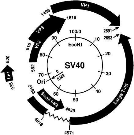
サウスカロライナ大学がん分子遺伝学教授のフィリップ・バックホーツ博士によれば、ファイザーのワクチンはプラスミドで "汚染 "されており、プラスミドがDNAに組み込まれ、ゲノムを改変した疑いがあるという16。
私はここで質問し、いくつかの点をつないでいるだけである。今のところ、癌を引き起こすプラスミドが悪意を持って意図的に挿入されたという証拠はない。今のところ、それはコンタミネーションであると主張されている。
癌を爆発的に増加させる原因は他にあるのだろうか?
コビッドワクチンに含まれる酸化グラフェンは、数時間以内に体内の抗酸化物質であるグルタチオンとSODを枯渇させる巨大なフリーラジカルであり、染色体を破壊し、DNAを切断し変異させ、細胞膜を切断し、大規模な炎症を引き起こす。DNAの突然変異が細胞をガン化させる主な原因であることは周知の通りだ。
細胞膜も破壊され、機能不全に陥ることで、ガンを引き起こす。その仕組みを説明しよう: ジェリー・テナント医学博士の著書『ヒーリング・イズ・ボルテージ』とロバート・O・ベッカー医学博士の著書『ボディ・エレクトリック』の情報17を結びつけると、細胞の電圧をプラスに変えると、細胞は成体幹細胞へと退行/脱分化することがわかる。
ベッカー博士は、サンショウウオが欠損した四肢を再生する際に細胞レベルで何が起こるかを調べるため、サンショウウオについて多くの研究を行った。博士は、哀れなサンショウウオの四肢を切断する前と後の健康な四肢細胞の細胞電圧を測定した。その結果、通常の電圧は-10mVであることがわかった。しかし、四肢を切断すると、電圧は+25mVまで上昇し、正常な細胞は成体幹細胞へと脱分化し、そこから新しい四肢組織が成長した。その後、新しい脚が成長する間に電圧は-30mVまで下がる。その間に電圧は徐々に下がり、通常の-10mVになる。つまり、電荷のマイナスからプラスへの反転に伴う電圧の上昇が、正常な組織細胞を成体幹細胞へと脱分化させる刺激となるようだ。
テナント博士はこの情報を基に、癌細胞と妊婦の胎盤細胞が基本的に同じ反応を示すことを発見した。両者とも他の組織に侵入し、血管新生を通じて自らの血液供給を組織化する。顕微鏡で見ると、胎盤細胞と癌細胞はよく似ており、どちらも絨毛性性腺刺激ホルモンを分泌する。つまり癌とは、身体が間違った場所とタイミングで胎盤を作ることに他ならない。胎盤を作る刺激とは、細胞がプラスの電圧に上昇することである。人間の正常な細胞電圧は-20~-25ミリボルトである。細胞が自己修復を必要とするとき、電圧は-50ミリボルトにさえなる。 テナント博士は、電圧が+25~+30ミリボルトよりさらに上昇すると、成体幹細胞は脱分化を続け、癌細胞になると理論化している。 電圧を-60mVという低電圧に戻すのに十分な電子を挿入すると、癌細胞は成体幹細胞に分化し、正常な組織細胞に戻るはずである。
彼は、電子泥棒を排除し、電子供与体を適用することによって、細胞をマイナス電圧の状態に戻すことができることを発見した。彼は、鍼治療の経絡と、バッテリーパックとして機能する特定の筋肉群や、特定の経絡につながった組織で電子の損失を引き起こす歯の感染症との関連についての情報を提供している。この記事の目的に最も関連する発見は、プラス電圧は組織幹細胞の脱分化によって細胞を癌細胞に変えるというものである。
では、何が細胞の電圧をマイナスに保つのだろうか?ナトリウム・カリウムポンプが細胞膜に埋め込まれている。 このポンプはプラスに帯電した3つのナトリウムイオンを細胞外に送り出し、マイナスに帯電した2つのカリウムイオンを細胞内に取り込む。これによって細胞の電圧はマイナスに保たれる。細胞膜が損傷すると、このポンプがその役割を果たせなくなり、細胞内の電圧はプラスに上昇する。
慢性的な炎症18も、細胞が癌化する原因と考えられているが、これは炎症によって傷ついた細胞膜のナトリウム・カリウムポンプの誤作動と関係があるかもしれない。酸化グラフェンは大量の炎症19を引き起こし、サイトカインIL-6、IL-12、TNFa、NFkbなどの炎症マーカーを上昇させる。
酸化グラフェンは免疫細胞、特に腫瘍の除去に関与する主要な細胞であるT1ヘルパー細胞とT2ヘルパー細胞の破壊を引き起こす。
自己組織化し、複製するナノテク・センサーとトランスミッター20は、5G経由で信号を送受信する能力を持つことが、ジャブジャブ生血液暗視野顕微鏡検査で、ジャブジャブ生とそうでない人の血液から発見されている。EMF信号を送受信するものが体内に存在することは、健康への悪影響という点で良いことではない。携帯電話と脳腫瘍の関連性は確立されている。このように、生体組織に近接したEMFには発がん作用がある。このようなナノデバイスは体内のいたるところにあり、常に信号を受発信している以上、発がん性の影響を及ぼさずにはいられない。
コビッドワクチンに含まれる発癌性物質に加え、私たちは日々、より多くの環境毒素にさらされている。これらは常に癌の主な原因となっている。2016年に出版した拙著『Meet Your Killers21』では、環境毒素とその健康への影響について600ページ以上にわたって詳しく述べている。
2015年に最も多かった癌は、乳癌、肺癌、気管支癌、前立腺癌、結腸・直腸癌、膀胱癌などと予測されている。最も多い癌のリストを見ると、最も影響を受ける臓器は、肺、結腸、直腸、膀胱、皮膚、腎臓のように、毒素に直接さらされるか、身体の解毒に関与していることがわかる。乳房や前立腺の組織は、プラスチック(ビスフェノールA)やフタル酸エステルから過剰に分泌されるエストロゲンや、前立腺癌の危険因子であるテストステロン不足などのホルモン不足など、ホルモンの変化に非常に敏感である。
脳腫瘍は、20歳未満の子供の癌死亡原因の第1位であり、20歳から39歳の若年成人の死亡原因の第3位である。携帯電話の放射線と関係があるのだろうか?
私たちの環境に存在する膨大な数の発癌性物質が、癌の発生に関係していることは明らかだ。既知の発癌物質はたくさんある。しかし、これらの発癌性物質はすべて、私たちを発癌性物質から守ってくれるはずの政府機関によって認可されているか、あるいは認可されていたものなのだ。以下は、私たちの環境に存在する一般的な発がん性物質のリストである:
発癌性物質:22
塩化ビニル
ダイオキシン類
六価クロム化合物
ビスフェノールA(BPA)
ベンゼン
アスベスト
ヒ素
加熱アスパルテーム由来のジケトピペラジン
ニトロソアミン
ブルーNr
シトラスレッドNr 2
イエローNr 6
臭素酸カリウム
グリホサート (ラウンドアップ®)
農薬中のヨウ化メチル
塩素副産物
パーソナルケア製品に含まれるジエタノールアミンのうちニトロソジエタノールアミン
ホルムアルデヒド
1,4 ジオキサン
コールタール
エチレンオキシド
ハイドロキノン
フェニレンジアミン
ラウリル硫酸ナトリウム
ブチルヒドロキシアニソール
日焼け止めに含まれるパルミチン酸レチニル
日焼け止めに含まれるパラアミノ安息香酸(PABA)
ベンゼン
アセトアルデヒド
プロピレングリコール
パラフィン
ある強度の電磁場、マイクロ波放射
化学物質の組み合わせは、互いの影響を増幅する可能性があります。少量の化学物質であっても、組み合わせると互いの悪影響を増幅させる。発癌性のない化学物質でさえ、協調して作用して癌を発生させることがある。28カ国の174人の科学者からなるタスクフォース23は、ヒトに対して発癌性がないと考えられている85の原型的化学物質を調査し、発癌に重要なメカニズムの長いリストに照らし合わせてその影響を検討した。
様々な癌の特徴に焦点を当てたチームに分かれ、研究グループは、調査した化学物質のうち50種が、ヒトが日常的に暴露しているレベルで、主要な癌関連メカニズムを支持していることを発見した。
この発見は、低レベルの化学物質への暴露では発癌性がなくても、化学物質が互いに作用し合って癌を引き起こす可能性があるという考えを支持するものである。
毒素を吸い込んだり、飲んだり、食べたり、皮膚から吸収したりすることは十分に悪いことだが、注射することより悪いことはない。
要約すると、SV40はウイルスではないが、ベロ細胞培養物から自然に、あるいは意図的に作られたプラスミド(環状DNA)であり、腫瘍抑制遺伝子をブロックするタンパク質を作り出す可能性がある。さらに、酸化グラフェンは、体内でナノテクノロジーから発生するEMFと同様に、主要な発癌性物質である。これは、環境中の既知の発癌性物質にさらされる機会が増えることに加えて起こる。
引用文献
1.SV40配列の存在に関するポリオワクチンの検査
D V Sangar 1、D J Wood、P D Minor
https://pubmed.ncbi.nlm.nih.gov/9776243/
2. ポリオワクチン製剤のSV40配列の検査
D Sangar 1、P A Pipkin、D J Wood、P D Minor
PMID: 10441397
DOI: 10.1006/biol.1998.0170
https://pubmed.ncbi.nlm.nih.gov/10441397/
3. ウイルス学
第24巻、第3号、1964年11月、381-387ページ
シミアンウイルス40の精製
著者リンク オーバーレイパネルを開く P.H. Black , E.M. Crawford , L.V. Crawford
https://doi.org/10.1016/0042-6822(64)90175-8
4. ウイルス学
第17巻、第1号、1962年5月、65-75ページ
アカゲザル腎臓細胞培養における発癌物質がシミアンウイルス40であることの同定
著者リンク オーバーレイパネルを開く バーニス・E・エディ、ジェラルド・S・ボーマン、ジョージ・E・グラブス、ラルフ・D・ヤング
https://doi.org/10.1016/0042-6822(62)90082-XGet
5. 実験分子病理学
第1巻、第5号、1962年10月、397-416ページ
組織培養における類人猿空胞化ウイルス(SV-40)の増殖に関する免疫蛍光学的、細胞化学的および微小細胞学的研究
著者リンク オーバーレイパネルを開くHeather Donald Mayor, Sara E. Stinebaugh, Richard M. Jamison, Liane E. Jordan, Joseph L. Melnick
https://doi.org/10.1016/0014-4800(62)90033-3
6. ウイルス学
第16巻、第3号、1962年3月、348-350ページ
考察と予備報告
空胞化ウイルスSV40によるプラーク形成
著者リンク オーバーレイパネルを開くSara Stinebaugh, Joseph L. Melnick
https://doi.org/10.1016/0042-6822(62)90259-3:
7. ウイルス学
第16巻、第3号、1962年3月、325-333ページ
サル腎臓組織培養細胞におけるポリオウイルスの形成
著者リンク オーバーレイパネルHeather Donald Mayor, Liane E. Jordan
https://doi.org/10.1016/0042-6822(62)90254-4
8. ヒト癌におけるSV40の役割はあるか?
Danielle L Poulin 1, James A DeCaprio
PMID: 16963733
論文番号:10.1200/jco.2005.03.7101
https://pubmed.ncbi.nlm.nih.gov/16963733/
9. ウイルス学
第17巻、第1号、1962年5月、65-75ページ
アカゲザル腎臓細胞培養における発癌物質がシミアンウイルス40であることの同定
バーニス・E・エディ、ジェラルド・S・ボーマン、ジョージ・E・グラブス、ラルフ・D・ヤング
https://doi.org/10.1016/0042-6822(62)90082-X
10. Clin Microbiol Rev. 2004 Jul; 17(3): 495-508.
doi: 10.1128/CMR.17.3.495-508.2004
PMCID: PMC452549 PMID: 15258090
ヒトの新たな病原体シミアンウイルス40とがんにおけるその役割
レジス・A・ヴィルチェス1,2およびジャネット・S・ビュテル2,*。
11.ネイチャー
公開日:2015年09月03日
癌におけるLINE-1:多面的機能と臨床的意義の可能性
Lu Xiao-Jie MD、Xue Hui-Ying、Xiaolong Qi MD、Xu Jiang & Ma Shi-Jie
Genetics in Medicine volume 18, pages431-439 (2016) https://www.nature.com/articles/gim2015119#:~:text=Long%20interspersed%20nuclear%20element%2D1,intermediates%2C%20a%20process%20termed%20retrotransposition.
12. ヒトゲノムにおける高親和性SV40 T抗原結合部位
クラウディア・グルース1、エラ・ヴェッツェル2、マルティナ・バーク、ウルスラ・モック3、ロルフ・クニッパーズ
https://doi.org/10.1016/0042-6822(88)90095-5
https://www.sciencedirect.com/science/article/abs/pii/0042682288900955
13. アカゲザルはヒト側枝腎形成のモデルである
Schuh, Meredith P.; Alkhudairy, Lyan; Potter, Andrew; Potter, S. Steven; Chetal, Kashish; Thakkar, Kairavee; Salomonis, Nathan; Kopan, Raphael
JASN 32(5):p 1097-1112, May 2021. | 論文|doi: 10.1681/asn.2020101459
https://journals.lww.com/jasn/fulltext/2021/05000/The_Rhesus_Macaque_Serves_As_a_Model_for_Human.15.
14. ロタウイルスにコードされた非構造タンパク質1は、腫瘍抑制タンパク質p53を標的として細胞アポトーシス機構を調節する
著者 Rahul Bhowmick, Umesh Chandra Halder, Shiladitya Chattopadhyay, Mukti Kant Nayak, Mamta Chawla-SarkarAUTHORS INFO & AFFILIATIONS
DOI: https://doi.org/10.1128/jvi.00734-13
https://journals.asm.org/doi/full/10.1128/jvi.00734-13
15. https://blog.genofab.com/vero-cells
16. chrome-extension://efaidnbmnnibpcajpcglclefindmkaj/https://www.scstatehouse.gov/CommitteeInfo/SenateMedicalAffairsCommittee/PandemicPreparedness/Phillip-Buckhaults-SC-Senate-09122023-final.pdf プラズマミド汚染
17. https://www.plaquex.com/_files/ugd/2fa22c_e42f109fa88e4a868bb305aa9fb30128.pdf
18. Immunity. 2019 Jul 16; 51(1): 27-41. doi: 10.1016/j.immuni.2019.06.025 PMCID: PMC6831096 NIHMSID: NIHMS1533333 PMID: 31315034
炎症とがん: トリガー、メカニズム、結果
フロリアン R. グレーテン、セルゲイ I. グリヴェンニコフ
https://www.ncbi.nlm.nih.gov/pmc/articles/PMC6831096/
19. グラフェンファミリーナノ粒子の毒性:起源とメカニズムの総説。Particle and Fibre Toxicology (216) 13:57 DOI 10.1186/s12989-016-0168-y Lingling Ou et al.
20. https://karenkingston.substack.com/p/addressing-the-threat-of-synthetic
21. https://www.lulu.com/shop/anita-baxas-md/meet-your-killers/paperback/product-1z47mg5d.html?q=anita+baxas&page=1&pageSize=4
22. 既知の発癌物質: www.psr.org/environment-and-health/confronting- toxics/examples-of-environmental-carcinogens.html
23. https://www.sciencedaily.com/releases/2015/06/150623072237.htm
出典:
アニタ・バクサス教授 - サブスタック - https://anitabaxasmd.substack.com
丁寧なお知らせ - 私は、イェードン博士を取り上げた私の記事に対する嫌がらせ、侮辱、罵倒、罵詈雑言、嘘、そして私の回答(および賛同者の回答)、さらには「モデレーター」であるローダ・ウィルソンによる記事全体の検閲・削除を受け、またしてもExpose Telegramグループから追放された。私はこの件に関して継続的に助けを求めてきたが、それでもまだ続いている......自分ではどうしようもないいじめを受け続けることはできない。従って、以下の記事は通常のように自動的に投稿されることはありません。ですから、どうか心ある読者の皆さんには、人類に対する犯罪を暴くことによって、私たち全員のために本当に働いてくれている目覚めた研究者、科学者、医師たちの重要な仕事を共有する手助けをしていただけないでしょうか?TIA
スペイン語訳:
La historia del cáncer SV40 cuestionada - No es un virus, pero ¿cuál es la conexión con el cáncer?
POR PATRICIA HARRITY
30 DE ABRIL DE 2024
Durante las últimas décadas se ha afirmado que las infecciones por el virus simio 40 (SV40) causan cáncer en los seres humanos. Esto se debe principalmente a que la secuencia genética atribuida al SV40 se ha encontrado en varias células cancerosas de seres humanos y animales de laboratorio. Según la doctora en medicina profesora Anita Baxas, esto se debe a que "los animales de laboratorio desarrollaron cáncer cuando se les inyectó un brebaje lanzado en los cultivos de células Vero que decía contener SV40". Sin embargo, el SV40 no es un virus, sino que puede tratarse de un plásmido (ADN circular) construido a propósito o de forma natural a partir de cultivos de células Vero que crea una proteína que bloquea los genes supresores de tumores, afirma la profesora Baxas.
El profesor también afirma que "los mismos procedimientos para la detección de virus se han hecho para SV40 y no proporciona ninguna prueba de que es un "virus", ni que incluso existe ver aquí La teoría del virus cuestionable
En el siguiente artículo el profesor Baxas Cuestiona la historia del cáncer sv40 y explica que los principales carcinógenos como el óxido de grafeno y EMFs son recibidos y emitidos por la nano tecnología dentro del cuerpo y junto con la exposición aprobada por el gobierno a carcinógenos conocidos en el medio ambiente es lo que realmente podría ser lo que está causando la "explosión del cáncer.'
La historia del cáncer SV40 cuestionada.
por Anita Baxas MD
Durante casi 60 años se ha afirmado que las infecciones por el virus Simian 40 causan cáncer en los seres humanos. El principal sospechoso ha sido la vacuna contra la polio1,2 ampliamente utilizada a finales de los años 50 cultivada en cultivos de células de riñón de mono verde, las células Vero que todavía se utilizan hoy en día como cultivos celulares para "probar" la existencia de "virus" y cultivarlos.
Existe una amplia falta de pruebas de que los virus existan. Para no extender demasiado este artículo, lea el Substack que explica esta falta de pruebas aquí: https://anitabaxasmd.substack.com/p/the-questionable-virus-theory.
Como para el SV40 se han realizado los mismos procedimientos,3,4,5,6,7 de detección de virus que para todos los demás "virus", esto tampoco aporta ninguna prueba de que el SV40 exista y sea un virus. La principal razón por la que se le acusa de causar cáncer es que la secuencia genética atribuida al SV40 se ha encontrado en diversas células cancerosas de seres humanos y animales de laboratorio8,9. Otra razón es que cuando a los animales de laboratorio se les inyectó un brebaje del material arrojado en los cultivos de células Vero que supuestamente contienen SV40, desarrollaron cánceres. El mecanismo alegado es que el SV40 inhibe ciertos genes que supuestamente suprimen el crecimiento tumoral. Se trata de genes supresores de tumores como el p53 y otros10. La razón es que el virus quiere que la célula replique el virus y, por lo tanto, necesita detener los procesos que lo inhibirían. Esto se atribuye a un antígeno T que se une a estos genes supresores y detiene su función supresora de la división celular11..
¿Qué es un antígeno T? Es simplemente una secuencia genética entre la cadena de ADN atribuida al SV40 que codifica para una proteína que parece unirse a los genes supresores de tumores. El Antígeno T del SV40 parece unirse preferentemente al ADN L-1 de las células humanas. Estos también se denominan genes saltarines que utilizan un mecanismo de copia-pega para propagarse por todo el genoma. Los L-1 están activos en la línea germinal durante la formación de un embrión. En el crecimiento del cáncer se activan erróneamente y pueden causar inestabilidad en el genoma, un sello distintivo para la aparición de la división celular incontrolada, también conocida como cáncer12.
¿Qué es un tumor? Básicamente es la proliferación de células por mitosis (división celular). Esto es lo que ocurre, por ejemplo, cuando un feto crece durante el embarazo. Durante este tiempo, estos genes supresores de tumores no están activos, o las células no podrían dividirse y el feto no podría desarrollarse y crecer.
¿Podría haber algo en los cultivos de células renales de mono que pudiera tener este efecto?
Un estudio no relacionado examinó el mecanismo por el que determinadas células renales13 (nefronas) se multiplican durante el último trimestre del embarazo. Para que estas células renales concretas se multipliquen se requiere una secuencia genética concreta. Estas secuencias genéticas codifican proteínas que inhiben los genes supresores de tumores. El término gen supresor de tumores es un poco engañoso. Se trata simplemente de un gen que sirve para inhibir la división celular cuando es apropiado hacerlo. Durante el crecimiento del tejido fetal tal inhibición no es apropiada, ya que el feto crece por división celular. Examinaron el mecanismo en riñones de fetos de mono del mismo tipo de mono del que derivan las células renales de mono (células Vero). Descubrieron que el mecanismo en humanos y en estos monos es idéntico. ¿Podrían ser estas secuencias genéticas las malinterpretadas como las del SV40? Parece que también se acusa a otros "virus" de inhibir genes supresores de tumores, como el Rotavirus14. Estos también se habrían "encontrado" en cultivos de células Vero.
En 2014 investigadores examinaron los cultivos de células Vero15 y la secuencia genética. Entre otras anomalías encontraron una importante deleción del cromosoma 12 que contiene genes importantes para las funciones del ciclo celular. Esto podría estar detrás de la replicación continua de las células Vero en cultivo. Así, estas células se dividen y replican constantemente. Si las vacunas se fabrican a partir de este brebaje de cultivo celular y suero fetal bovino (¡FETAL!), ¿es posible que el impulso de replicación se transfecte a células humanas que luego comienzan a replicarse en un tumor?
¿Podría haber otra explicación?
El ADN del supuesto SV40 es circular como un plásmido. Los plásmidos son moléculas de ADN circulares fuera del cromosoma. A menudo se construyen e insertan en bacterias E. Coli o en cultivos celulares para que produzcan una proteína específica. Ahora podríamos volvernos un poco paranoicos y pensar que tal vez, por razones nefastas, se fabricaron plásmidos y se insertaron en los cultivos de células Vero utilizados para crear vacunas o directamente en las "vacunas", sobre todo en las más recientes, para crear a propósito una epidemia de cáncer.
ADN circular del SV40
Según Phillip Buckhaults, Doctor y Profesor de Genética Molecular del Cáncer de la Universidad de Carolina del Sur, las vacunas de Pfizer estaban "contaminadas" con plásmidos, y sospecha que se integraron en el ADN y modificaron el genoma.16
Sólo estoy haciendo preguntas y conectando algunos puntos aquí. Hasta ahora no hay pruebas de la inserción intencionada y maliciosa de plásmidos cancerígenos. Por ahora se afirma que fue una contaminación.
¿Qué otra cosa podría estar causando la explosión de cánceres?
El óxido de grafeno que se encuentra en las inyecciones de Covid es un radical libre masivo que agota los antioxidantes del cuerpo, el glutatión y el SOD, en pocas horas, dejándolo indefenso frente a este monstruoso radical libre, que puede desbocarse y destruir cromosomas, romper y mutar el ADN, cortar las membranas celulares y provocar una inflamación masiva. Todos sabemos que la mutación del ADN es una de las principales causas de que las células se vuelvan cancerosas.
La destrucción y el mal funcionamiento de las membranas celulares también causan cáncer. Permítanme explicar cómo funciona esto: Cuando conectamos la información17 del Dr. Jerry Tennant, MD en su libro llamado Healing is Voltage y del Dr. Robert O. Becker, MD en su libro The Body Electric encontramos que cambiar el voltaje de una célula a positivo causa que la célula retroceda/desdiferencie a una célula madre adulta.
El Dr. Becker investigó mucho con salamandras para ver qué ocurre a nivel celular cuando vuelven a crecer las extremidades que les faltan. Midió el voltaje celular de las células del miembro sano antes y después de amputar un miembro a las pobres salamandras. Descubrió que el voltaje normal era de - 10 mV. Pero después de amputar un miembro, el voltaje se disparaba a + 25 mV, lo que provocaba que las células normales se desdiferenciaran en células madre adultas de las que crecía el nuevo tejido del miembro. A continuación, el voltaje baja a -30 mV mientras crece una nueva pierna. Durante ese tiempo, el voltaje desciende gradualmente hasta los -10 mV normales. Así pues, parece que el aumento del voltaje con inversión de la carga de negativa a positiva es el estímulo que hace que las células de tejido normal se desdiferencien en células madre adultas.
El Dr. Tennant se basó en esta información y descubrió que las células cancerosas y las células de la placenta de las mujeres embarazadas reaccionan básicamente de la misma manera. Ambas invaden otros tejidos y organizan su propio riego sanguíneo mediante angiogénesis. Microscópicamente, las células placentarias y las células cancerosas se parecen, y ambas segregan la hormona gonadotrófica coriónica. Así pues, el cáncer no es más que el cuerpo fabricando una placenta en el lugar y el momento equivocados. El estímulo para hacer una placenta es el aumento celular a un voltaje positivo. El voltaje celular normal en los humanos es de -20 a -25 milivoltios. Cuando la célula necesita repararse, el voltaje llega incluso a -50mV. El Dr. Tennant teoriza que si el voltaje aumenta más allá de +25 a +30mV las células madre adultas continúan desdiferenciándose para convertirse en células cancerosas. Cuando se insertan suficientes electrones para que el voltaje vuelva a ser tan bajo como -60 mV, las células cancerosas deberían volver a diferenciarse en células madre adultas y luego en células de tejido normal.
Descubrió que se podía devolver a las células a un estado de voltaje negativo eliminando los ladrones de electrones y aplicando donantes de electrones. Proporciona información sobre la conexión entre los meridianos de acupuntura y grupos musculares específicos que actúan como baterías e infecciones dentales que causan la pérdida de electrones en los tejidos conectados al meridiano específico. Aunque fascinante, no voy a entrar en toda la información que proporciona como el hallazgo más relevante para el propósito de este artículo es que el voltaje positivo convierte las células en células cancerosas por desdiferenciación de las células madre del tejido.
Ahora bien, ¿qué es lo que mantiene negativo el voltaje de una célula? La bomba de sodio-potasio está incrustada en la membrana celular. Bombea tres iones de sodio cargados positivamente fuera de la célula y deja entrar dos iones de potasio cargados negativamente. De este modo, el voltaje de la célula se mantiene negativo. Cuando la membrana celular está dañada, esta bomba no puede hacer su trabajo y el voltaje dentro de la célula sube a una carga positiva.
También se cree que la inflamación crónica18 es una de las razones por las que las células se vuelven cancerosas, lo que podría tener que ver con el mal funcionamiento de las bombas de sodio-potasio en las membranas celulares dañadas por la inflamación. El óxido de grafeno provoca una inflamación masiva19, elevando ciertos marcadores inflamatorios como las citocinas IL-6, IL-12, TNFa y NFkb.
El óxido de grafeno provoca la destrucción de las células inmunitarias, en particular de las células T1 y T2 auxiliares, que son las principales células implicadas en la eliminación de tumores.
Se han hallado sensores y transmisores nanotecnológicos autoconstruibles y replicables20 en la sangre de personas inyectadas y no inyectadas en microscopía de campo oscuro de sangre viva que tienen la capacidad de recibir y enviar señales a través de 5G. Tener algo dentro del cuerpo que recibe y envía señales EMF seguramente no puede ser algo bueno en lo que respecta a los efectos negativos para la salud. Se ha establecido una relación entre los teléfonos móviles y los tumores cerebrales. Por lo tanto, los CEM cerca de tejidos vivos tienen efectos cancerígenos. Estos nanodispositivos están por todas partes dentro del cuerpo y no pueden sino tener efectos cancerígenos cuando están constantemente recibiendo y emitiendo señales.
Además de los carcinógenos de las inyecciones Covid, cada día estamos expuestos a más toxinas ambientales. Éstas siempre han sido una de las principales causas de cáncer. En mi libro, Meet Your Killers21, de 2016 elaboro más de 600 páginas sobre las toxinas ambientales y sus efectos en nuestra salud.
Los cánceres más comunes en 2015 se proyectaron como cáncer de mama, cáncer de pulmón y bronquios, cáncer de próstata, cáncer de colon y recto, cáncer de vejiga y más. Si observamos la lista de los cánceres más comunes, veremos que los órganos más afectados están expuestos directamente a las toxinas o participan en la desintoxicación del organismo, como los pulmones, el colon, el recto, la vejiga, la piel y los riñones. Los tejidos de la mama y la próstata son muy sensibles a los cambios hormonales, como el exceso de estrógenos procedentes de los plásticos (bisfenol A) y los ftalatos, o a la falta de hormonas, como la falta de testosterona, que es un factor de riesgo del cáncer de próstata.
El cáncer cerebral es la principal causa de muerte por cáncer en niños menores de veinte años y la tercera causa de muerte en adultos jóvenes de entre veinte y treinta y nueve años. ¿Podría tener algo que ver con la radiación de los teléfonos móviles?
Está claro que el enorme número de carcinógenos que hay en nuestro entorno tiene algo que ver con el desarrollo del cáncer. Hay muchos carcinógenos conocidos. Lo que debería hacerte hervir de rabia es que todos estos carcinógenos son o fueron aprobados en algún momento por una agencia gubernamental que se supone que debe protegernos de ellos. He aquí una lista de carcinógenos comunes en nuestro entorno:
Cancerígenos:22
Cloruro de vinilo
Dioxinas
Compuestos hexavalentes de cromo
Bisfenol A (BPA)
Benceno
Amianto
Arsénico
Diketopiperazina del aspartamo calentado
Nitrosamina
Azul Nr
Rojo cítrico Nr 2
Amarillo Nr 6
Bromato de potasio
Glifosato (Roundup®)
Yoduro de metilo en pesticidas
Subproductos del cloro
Nitrosodietanolamina a partir de dietanolamina en productos de cuidado personal
Formaldehído
1,4 Dioxano
Alquitrán de hulla
Óxido de etileno
Hidroquinona
Fenilendiamina
Lauril sulfato sódico
Butilhidroxianisol
Palmitato de retinol en protección solar
Ácido paraaminobenzoico (PABA) en los protectores solares
Benceno
Acetaldehído
Propilenglicol
Parafina
Campos electromagnéticos de cierta intensidad, radiación de microondas
Las sustancias químicas combinadas pueden amplificar sus efectos. Incluso pequeñas cantidades de sustancias químicas pueden amplificar sus efectos adversos cuando se combinan. Incluso las sustancias químicas no cancerígenas pueden actuar conjuntamente para producir cáncer. Un grupo de 174 científicos de 28 países23 investigó 85 sustancias químicas prototípicas que no se consideraban carcinógenas para los seres humanos, y revisaron sus efectos comparándolos con una larga lista de mecanismos que son importantes para el desarrollo del cáncer.
Trabajando en equipos que se centraron en varios rasgos distintivos del cáncer, el grupo descubrió que 50 de esas sustancias químicas examinadas apoyaban mecanismos clave relacionados con el cáncer a niveles a los que los seres humanos están expuestos de forma rutinaria.
El hallazgo respalda la idea de que las sustancias químicas pueden actuar conjuntamente para provocar cáncer, aunque la exposición a niveles bajos de estas sustancias por separado no sea cancerígena.
Respirar, beber, ingerir toxinas y absorberlas a través de la piel ya es bastante malo, pero no puede ser mucho peor que recibirlas inyectadas.
En resumen, el SV40 no es un virus, sino que puede tratarse de un plásmido (ADN circular) natural de cultivos de células Vero o construido a propósito que crea una proteína que bloquea los genes supresores de tumores. Además, el óxido de grafeno es un carcinógeno importante, al igual que los CEM recibidos y emitidos por la nanotecnología dentro del cuerpo. Esto se suma al aumento de la exposición a carcinógenos conocidos en el medio ambiente.
Citas
1.Examen de vacunas antipoliomielíticas para detectar la presencia de secuencias de SV40
D V Sangar 1, D J Wood, P D Minor
https://pubmed.ncbi.nlm.nih.gov/9776243/
2. 2. Examen de preparaciones de vacunas de poliovirus para detectar la presencia de secuencias de SV40
D Sangar 1, P A Pipkin, D J Wood, P D Minor
PMID: 10441397
DOI: 10.1006/biol.1998.0170
https://pubmed.ncbi.nlm.nih.gov/10441397/
3. Virología
Volumen 24, Número 3, Noviembre 1964, Páginas 381-387
La purificación del virus simio 40
Los enlaces de los autores abren el panel superpuesto P.H. Black , E.M. Crawford , L.V. Crawford
https://doi.org/10.1016/0042-6822(64)90175-8
4. Virología
Volumen 17, Número 1, Mayo 1962, Páginas 65-75
Identificación de la sustancia oncogénica en cultivos de células renales de mono rhesus como virus simio 40
Los enlaces de los autores abren el panel superpuesto Bernice E. Eddy, Gerald S. Borman, George E. Grubbs, Ralph D. Young
https://doi.org/10.1016/0042-6822(62)90082-XGet
5. Patología Experimental y Molecular
Volumen 1, Número 5, Octubre 1962, Páginas 397-416
Estudios inmunofluorescentes, citoquímicos y microcitológicos sobre el crecimiento del virus vacuolador simio (SV-40) en cultivo de tejidos
Autor enlaces abrir ventana superpuestaHeather Donald Mayor, Sara E. Stinebaugh, Richard M. Jamison, Liane E. Jordan, Joseph L. Melnick
https://doi.org/10.1016/0014-4800(62)90033-3
6. Virología
Volumen 16, número 3, marzo de 1962, páginas 348-350
Discusión e informe preliminar
Formación de placas por el virus vacuolador, SV40
Enlaces del autor abrir panel superpuestoSara Stinebaugh, Joseph L. Melnick
https://doi.org/10.1016/0042-6822(62)90259-3:
7. Virología
Volumen 16, Número 3, Marzo 1962, Páginas 325-333
Formación de poliovirus en células de cultivo de tejidos de riñón de mono
Los enlaces del autor abren el panel superpuestoHeather Donald Mayor, Liane E. Jordan
https://doi.org/10.1016/0042-6822(62)90254-4
8. ¿Existe un papel para el SV40 en el cáncer humano?
Danielle L Poulin 1, James A DeCaprio
PMID: 16963733
DOI: 10.1200/JCO.2005.03.7101
https://pubmed.ncbi.nlm.nih.gov/16963733/
9. Virología
Volumen 17, Número 1, Mayo 1962, Páginas 65-75
Identificación de la sustancia oncogénica en cultivos de células renales de mono rhesus como virus simio 40
Bernice E. Eddy, Gerald S. Borman, George E. Grubbs, Ralph D. Young
https://doi.org/10.1016/0042-6822(62)90082-X
10. Clin Microbiol Rev. 2004 Jul; 17(3): 495-508.
doi: 10.1128/CMR.17.3.495-508.2004
PMCID: PMC452549 PMID: 15258090
Patógeno humano emergente Virus Simian 40 y su papel en el cáncer
Regis A. Vilchez1,2 y Janet S. Butel2,*
11. nature
Publicado: 03 septiembre 2015
LINE-1 en el cáncer: funciones multifacéticas y posibles implicaciones clínicas
Dr. Lu Xiao-Jie, Dr. Xue Hui-Ying, Dr. Xiaolong Qi, Dr. Xu Jiang y Dr. Ma Shi-Jie
Genetics in Medicine volumen 18, páginas431-439 (2016) https://www.nature.com/articles/gim2015119#:~:text=Long%20interspersed%20nuclear%20element%2D1,intermediates%2C%20a%20process%20termed%20retrotransposition.
12. Sitios de unión del antígeno T del SV40 de alta afinidad en el genoma humano
Claudia Gruss 1, Ella Wetzel 2, Martina Baack, Ursula Mock 3, Rolf Knippers
https://doi.org/10.1016/0042-6822(88)90095-5
https://www.sciencedirect.com/science/article/abs/pii/0042682288900955
13. El macaco Rhesus sirve de modelo para la nefrogénesis de la rama lateral humana
Schuh, Meredith P.; Alkhudairy, Lyan; Potter, Andrew; Potter, S. Steven; Chetal, Kashish; Thakkar, Kairavee; Salomonis, Nathan; Kopan, Raphael
JASN 32(5):p 1097-1112, mayo de 2021. | DOI: 10.1681/ASN.2020101459
https://journals.lww.com/jasn/fulltext/2021/05000/The_Rhesus_Macaque_Serves_As_a_Model_for_Human.15.
14. 14. Rotavirus-Encoded Nonstructural Protein 1 Modulates Cellular Apoptotic Machinery by Targeting Tumor Suppressor Protein p53
Autores: Rahul Bhowmick, Umesh Chandra Halder, Shiladitya Chattopadhyay, Mukti Kant Nayak, Mamta Chawla-SarkarINFORMACIÓN DEL AUTOR Y AFILIACIONES
DOI: https://doi.org/10.1128/jvi.00734-13
https://journals.asm.org/doi/full/10.1128/jvi.00734-13
15. https://blog.genofab.com/vero-cells
16. chrome-extension://efaidnbmnnnibpcajpcglclefindmkaj/https://www.scstatehouse.gov/CommitteeInfo/SenateMedicalAffairsCommittee/PandemicPreparedness/Phillip-Buckhaults-SC-Senate-09122023-final.pdf CONTAMINACIÓN DEL PLÁSMIDO
17. https://www.plaquex.com/_files/ugd/2fa22c_e42f109fa88e4a868bb305aa9fb30128.pdf
18. Immunity. 2019 Jul 16; 51(1): 27-41. doi: 10.1016/j.immuni.2019.06.025 PMCID: PMC6831096 NIHMSID: NIHMS1533333 PMID: 31315034
Inflamación y cáncer: Desencadenantes, mecanismos y consecuencias
Florian R. Greten y Sergei I. Grivennikov
https://www.ncbi.nlm.nih.gov/pmc/articles/PMC6831096/
19. Toxicidad de las nanopartículas de la familia del grafeno: revisión general de los orígenes y mecanismos. Particle and Fibre Toxicology (216) 13:57 DOI 10.1186/s12989-016-0168-y Lingling Ou et al.
20. https://karenkingston.substack.com/p/addressing-the-threat-of-synthetic
21. https://www.lulu.com/shop/anita-baxas-md/meet-your-killers/paperback/product-1z47mg5d.html?q=anita+baxas&page=1&pageSize=4
22. Carcinógenos conocidos: www.psr.org/environment-and-health/confronting- toxics/examples-of-environmental-carcinogens.html
23. https://www.sciencedaily.com/releases/2015/06/150623072237.htm
Fuente:
Profesora Anita Baxas - Substack - https://anitabaxasmd.substack.com
Aviso cortés - Me han vuelto a expulsar del grupo Expose Telegram tras mi artículo sobre el Dr. Yeadon y meses de acoso, insultos, abusos, insultos, mentiras y censura/eliminación de mis respuestas (y de las de aquellos que me apoyan) e incluso de artículos enteros por parte de Rhoda Wilson, la "moderadora". No puedo seguir sometiéndome al acoso sin tener ningún control sobre ello. Por lo tanto, el siguiente artículo no se publicará automáticamente allí, como suele ser el caso. Por lo tanto, ¿puedo pedir a los lectores con ideas afines que ayuden a compartir el importante trabajo de los investigadores, científicos y médicos despiertos que realmente trabajan para todos nosotros sacando a la luz los crímenes contra la humanidad? TIA
原文:
The SV40 Cancer Story Questioned – It’s not a Virus, But What’s the Connection to Cancer?
BY PATRICIA HARRITY
ON APRIL 30, 2024
It has been claimed for the last few decades that Simian Virus 40 (SV40) infections cause cancer in humans. this is mainly because the genetic sequence attributed to SV40 has been found in various cancer cells in humans and lab animals. According to medical doctor Professor Anita Baxas this is because ‘lab animals developed cancer when injected with a concoction thrown into the Vero cell cultures claimed to contain SV40.’ However, SV40 is not a virus, but may well be plasmid (circular DNA) either purposely constructed or naturally from Vero cell cultures that creates a protein which blocks tumour suppressor genes, claims Professor Baxas.
The Professor also claims that ‘the same procedures for virus detection have been done for SV40 and provides no evidence that it is a ‘virus’ nor that it even exists see here The Questionable Virus Theory
In the following article Professor Baxas Questions the sv40 cancer story and explains that major carcinogens such as Graphene Oxide and EMFs are received and emitted by nano technology inside the body and along with government approved exposure to known carcinogens in the environment is what could actually be what is causing the “explosion of cancer.’
The SV40 Cancer Story Questioned.
by Anita Baxas MD
For nearly 60 years it has been claimed that Simian Virus 40 infections cause cancer in humans. The prime suspect has been the polio vaccine1,2 widely used in the late 1950ies grown in green monkey kidney cell cultures, the Vero cells still used today as cell cultures to “prove” the existence of and to grow “viruses”.
There is ample lack of evidence that viruses exist. To not overextend this article, please read the Substack explaining this lack of evidence here: https://anitabaxasmd.substack.com/p/the-questionable-virus-theory
As the same procedures,3,4,5,6,7 for virus detection have been done for SV40 as for all the other “viruses”, this too provides no evidence that SV40 exists and is a virus. The main reason why it is accused of causing cancer is that the genetic sequence attributed to SV40 has been found in various cancer cells in humans and lab animals8,9. Another reason is that when lab animals were injected with a concoction of the stuff thrown into the Vero cell cultures claimed to contain SV40, they develop cancers. The claimed mechanism is that SV40 inhibits certain genes that are supposed to suppress tumor growth. They are tumor suppressor genes such as p53 and others10. The reason is that the virus wants the cell to replicate the virus and thus needs to stop processes that would inhibit this. This is attributed to a T-Antigen that binds to these suppressor genes and thus stops their suppressing function of cell division11..
What is a T-Antigen? It’s simply a genetic sequence among the DNA strand attributed to SV40 that codes for a protein that seems to bind to tumor suppressor genes. The T Antigen of SV40 seems to bind preferably to L-1 DNA in human cells. These are also called jumping genes that use a copy – paste mechanism to propagate themselves throughout the genome. L-1 is active in the germ line during the formation of an embryo. In cancer growth they are erroneously activated and may cause instability in the genome, a hallmark for the emergence of uncontrolled cell division aka cancer12.
Now what is a tumor? It basically is the proliferation of cells by mitosis (cell division). This is what happens when a fetus grows during pregnancy for example. During this time, these tumor suppressor genes aren’t active, or cells couldn’t divide and the fetus couldn’t develop and grow.
Might there be something in monkey kidney cell cultures that can have this effect?
An unrelated study examined the mechanism by which particular kidney cells13 (nephrons) multiply during the last trimester of pregnancy. A particular genetic sequence is required in order for these particular kidney cells to multiply. These genetic sequences code for proteins that inhibit tumor suppressor genes. The term tumor suppressor gene is a bit misleading. It’s simply a gene that serves to inhibit cell division when it is appropriate to do so. During fetal tissue growth such inhibition is not appropriate as the fetus grows by cell division. They examined the mechanism in monkey fetus kidneys of the same type of monkey that the monkey kidney cells (Vero cells) are derived from. They found the mechanism in humans and these monkeys is identical. Could these genetic sequences be the ones misinterpreted as the ones from SV40? It seems other “viruses” are also accused of inhibiting tumor suppressor genes such as Rotavirus14. These too would have been “found” in Vero cell cultures.
In 2014 researchers examined Vero cell cultures15 and the genetic sequence. Among other abnormalities they found a major deletion of chromosome 12 that contains genes important for cell cycle functions. This could lie behind the continuous replication of Vero cells in culture. Thus, these cells constantly divide and replicate. If vaccines are made from this concoction of cell culture, fetal bovine serum (FETAL!) is it possible that the drive to replicate is transfected to human cells that then begin replicating into a tumor?
Could there be another explanation?
The DNA of the claimed SV40 is circular like a plasmid. Plasmids are circular DNA molecules outside of the chromosome. They are often constructed and inserted into E. Coli bacteria or cell cultures in order to have them produce a specific protein. Now we could become a little paranoid and think that maybe for nefarious reasons, plasmids were made and inserted into either the Vero cell cultures used to create vaccines or directly into “vaccines”, particularly more recent ones, to purposefully create an epidemic of cancer.
Circular DNA of SV40
According to Phillip Buckhaults, Ph.D. Professor of Cancer Molecular Genetics University of South Carolina, Pfizer jabs were “contaminated” with plasmids, and he suspects they were integrated into the DNA and modified the genome.16
I’m just asking questions and connecting some dots here. There is so far no evidence for the malicious purposeful insertion of cancer – causing plasmids. As of now it is claimed that it was a contamination.
What else could be causing the explosion of cancers?
Graphene Oxide found in the Covid jabs is a massive free radical that depletes the body’s antioxidants Glutathione and SOD within a few hours leaving it defenseless against this monstrous free radical which then can go on a rampage and destroy chromosomes, break and mutate DNA, cut up cell membranes and cause massive inflammation. We all know that mutating DNA is a major cause for cells to turn cancerous.
Destroyed and malfunctioning cell membranes too are causing cancer. Let me explain how that works: When we connect the information17 of Dr. Jerry Tennant, MD in his book called Healing is Voltage and of Dr. Robert O. Becker, MD in his book The Body Electric we find that changing the voltage of a cell to positive causes the cell to regress/de-differentiate into an adult stem cell.
Dr. Becker did a lot of research on Salamanders to see what happens on a cellular level when they regrow a missing limb. He measured the cellular voltage of healthy limb cells before and after amputating a limb from the poor Salamanders. He found that the normal voltage was – 10 mV. But after amputating a limb, the voltage shot up to + 25 mV which caused normal cells to dedifferentiate into adult stem cells out of which new limb tissue grew. Then the voltage drops to -30 mV while a new leg is grown. During that time the voltage gradually drops to the normal -10mV. So, it looks like the increase in voltage with reversal of the charge from negative to positive is the stimulus that causes normal tissue cells to dedifferentiate into adult stem cells.
Dr. Tennant built on this information and discovered that cancer cells and placental cells in pregnant women basically react the same way. Both invade other tissues and organize their own blood supply through angiogenesis. Microscopically placental cells and cancer cells look alike, and both secrete chorionic gonadotrophic hormone. So, cancer is nothing more than the body making a placenta in the wrong place and at the wrong time. The stimulus to make a placenta is the cellular increase to a positive voltage. Normal cellular voltage in humans is -20 to -25 millivolts. When the cell needs to repair itself, the voltage even goes to -50mV. Dr. Tennant theorizes that if the voltage increases further than +25 to +30mV the adult stem cells continue to dedifferentiate to become cancer cells. When you insert enough electrons to return the voltage to as low as -60mV, the cancer cells should differentiate back into adult stem cells and then back into normal tissue cells.
He discovered that you could return cells into a state of negative voltage by eliminating electron stealers and apply electron donors. He provides information on the connection between acupuncture meridians and specific muscle groups that act as battery packs and tooth infections that cause the loss of electrons on tissues connected to the specific meridian. Although fascinating, I won’t go into all the information he provides as the most relevant finding for the purpose of this article is that positive voltage turns cells into cancer cells by dedifferentiation of tissue stem cells.
Now what keeps a cells’ voltage negative? The Sodium Potassium pump is embedded in the cell membrane. It pumps out three positively charged Sodium ions out of the cell and lets in two negatively charged Potassium ions. This keeps the cells’ voltage negative. When the cell membrane is damaged, this pump can’t do its job and the voltage inside the cell climbs to a positive charge.
Chronic inflammation18 is also thought to be a reason that cells turn cancerous which might have to do with the malfunctioning sodium-potassium pumps in cell membranes damaged by inflammation. Graphene Oxide causes massive inflammation19, elevating certain inflammatory markers such as the Cytokines IL-6, IL-12, TNFa and NFkb.
Graphene Oxides causes destruction of immune cells, particularly of T1 and T2 Helper cells which are the primary cells involved in tumor elimination.
Self – constructing and replicating nanotech sensors and transmitters20 have been found in blood of the jabbed and unjabbed in live blood dark field microscopy that have the ability to receive and send signals via 5G. Having something inside the body that that receives and sends out EMF signals surely can’t be a good thing in regard to negative health effects. A link between cell phones and brain tumors has been established. Thus, EMF in close proximity to living tissue has carcinogenic effects. These nano devices are everywhere inside the body and cannot but have carcinogenic effects when they are constantly receiving and emitting signals.
In addition to the carcinogens in the Covid jabs, we are exposed daily by more and more Environmental Toxins. These have always been a major cause of cancers. In my book, Meet Your Killers21, from 2016 I elaborate on over 600 pages on environmental toxins and their effects on our health.
The most common cancers in 2015 were projected to be breast cancer, lung and bronchus cancer, prostate cancer, colon and rectum cancer, bladder cancer and more. If you look at the list of the most common cancers, you can see that the organs most affected are either directly exposed to toxins or are involved in detoxifying the body, like the lungs, colon, rectum, bladder, skin, and kidney. Breast and prostate tissues are very sensitive to hormonal changes such as too much estrogen from plastics (bisphenol A) and phthalates, or a lack of hormone such as a lack of testosterone, which is a risk factor for prostate cancer.
Brain cancer is the leading cause of cancer death in children under the age of twenty and the third leading cause of death in young adults ages twenty to thirty-nine. Might that have something to do with cell phone radiation?
Clearly, the huge number of carcinogens in our environment has something to do with the development of cancer. There are many known carcinogens. What should make you boiling mad is that all these carcinogens are or were approved by a government agency some time or another that is supposed to protect us from them. Here’s a list of common carcinogens in our environment:
Carcinogens:22
Vinyl Chloride
Dioxins
Chromium hexavalent compounds
Bisphenol A (BPA)
Benzene
Asbestos
Arsenic
Diketopiperazine from heated aspartame
Nitrosamine
Blue Nr
Citrus Red Nr 2
Yellow Nr 6
Potassium Bromate
Glyphosate (Roundup®)
Methyl iodide in pesticides
Chlorine by-products
Nitrosodiethanolamine out of diethanolamine in personal care products
Formaldehyde
1,4 Dioxane
Coal Tar
Ethylene Oxide
Hydroquinone
Phenylenediamine
Sodium Lauryl Sulfate
Butylated Hydroxyanisole
Retinyl Palmitate in sunscreen
Paraaminobenzoic Acid (PABA) in sunscreen
Benzene
Acetaldehyde
Propylene Glycol
Paraffin
Electromagnetic Fields of certain strength, Microwave Radiation
Chemicals in combination can amplify each other’s effects. Even small amounts of chemicals can amplify each other’s adverse effects when combined. Even noncarcinogenic chemicals can act in concert to produce cancer. A taskforce of 174 scientists in 28 countries23 investigated 85 prototypic chemicals that were not considered to be carcinogenic to humans, and they reviewed their effects against a long list of mechanisms that are important for cancer development.
Working in teams that focused on various hallmarks of cancer, the group found that 50 of those chemicals examined supported key cancer-related mechanisms at levels at which humans are routinely exposed.
The finding supports the idea that chemicals may be capable of acting in concert with one another to cause cancer, even though low-level exposures to these chemicals individually might not be carcinogenic.
Breathing in, drinking, eating toxins and absorbing them through the skin is bad enough, but it can’t get much worse than having them injected.
In summary SV40 is not a virus, but it may well be a plasmid (circular DNA) either naturally from Vero cell cultures or purposefully constructed that creates a protein which blocks tumor suppressor genes. In addition, Graphene Oxide is a major carcinogen as are EMFs received and emitted by nano technology inside the body. This is on top of the increased exposure to known carcinogens in the environment.
Citations
1.Examination of polio vaccines for the presence of SV40 sequences
D V Sangar 1, D J Wood, P D Minor
https://pubmed.ncbi.nlm.nih.gov/9776243/
2. Examination of poliovirus vaccine preparations for SV40 sequences
D Sangar 1, P A Pipkin, D J Wood, P D Minor
PMID: 10441397
DOI: 10.1006/biol.1998.0170
https://pubmed.ncbi.nlm.nih.gov/10441397/
3. Virology
Volume 24, Issue 3, November 1964, Pages 381-387
The purification of simian virus 40
Author links open overlay panel P.H. Black , E.M. Crawford , L.V. Crawford
https://doi.org/10.1016/0042-6822(64)90175-8
4. Virology
Volume 17, Issue 1, May 1962, Pages 65-75
Identification of the oncogenic substance in rhesus monkey kidney cell cultures as simian virus 40
Author links open overlay panel Bernice E. Eddy, Gerald S. Borman, George E. Grubbs, Ralph D. Young
https://doi.org/10.1016/0042-6822(62)90082-XGet
5. Experimental and Molecular Pathology
Volume 1, Issue 5, October 1962, Pages 397-416
Immunofluorescent, cytochemical, and microcytological studies on the growth of the simian vacuolating virus (SV-40) in tissue culture
Author links open overlay panelHeather Donald Mayor, Sara E. Stinebaugh, Richard M. Jamison, Liane E. Jordan, Joseph L. Melnick
https://doi.org/10.1016/0014-4800(62)90033-3
6. Virology
Volume 16, Issue 3, March 1962, Pages 348-350
Discussion and preliminary report
Plaque formation by vacuolating virus, SV40
Author links open overlay panelSara Stinebaugh, Joseph L. Melnick
https://doi.org/10.1016/0042-6822(62)90259-3:
7. Virology
Volume 16, Issue 3, March 1962, Pages 325-333
Formation of poliovirus in monkey kidney tissue culture cells
Author links open overlay panelHeather Donald Mayor, Liane E. Jordan
https://doi.org/10.1016/0042-6822(62)90254-4
8. Is there a role for SV40 in human cancer?
Danielle L Poulin 1, James A DeCaprio
PMID: 16963733
DOI: 10.1200/JCO.2005.03.7101
https://pubmed.ncbi.nlm.nih.gov/16963733/
9. Virology
Volume 17, Issue 1, May 1962, Pages 65-75
Identification of the oncogenic substance in rhesus monkey kidney cell cultures as simian virus 40
Bernice E. Eddy, Gerald S. Borman, George E. Grubbs, Ralph D. Young
https://doi.org/10.1016/0042-6822(62)90082-X
10. Clin Microbiol Rev. 2004 Jul; 17(3): 495–508.
doi: 10.1128/CMR.17.3.495-508.2004
PMCID: PMC452549 PMID: 15258090
Emergent Human Pathogen Simian Virus 40 and Its Role in Cancer
Regis A. Vilchez1,2 and Janet S. Butel2,*
11. nature
Published: 03 September 2015
LINE-1 in cancer: multifaceted functions and potential clinical implications
Lu Xiao-Jie MD, Xue Hui-Ying, Xiaolong Qi MD, Xu Jiang & Ma Shi-Jie
Genetics in Medicine volume 18, pages431–439 (2016) https://www.nature.com/articles/gim2015119#:~:text=Long%20interspersed%20nuclear%20element%2D1,intermediates%2C%20a%20process%20termed%20retrotransposition.
12. High-affinity SV40 T-antigen binding sites in the human genome
Claudia Gruss 1, Ella Wetzel 2, Martina Baack, Ursula Mock 3, Rolf Knippers
https://doi.org/10.1016/0042-6822(88)90095-5
https://www.sciencedirect.com/science/article/abs/pii/0042682288900955
13. The Rhesus Macaque Serves As a Model for Human Lateral Branch Nephrogenesis
Schuh, Meredith P.; Alkhudairy, Lyan; Potter, Andrew; Potter, S. Steven; Chetal, Kashish; Thakkar, Kairavee; Salomonis, Nathan; Kopan, Raphael
JASN 32(5):p 1097-1112, May 2021. | DOI: 10.1681/ASN.2020101459
https://journals.lww.com/jasn/fulltext/2021/05000/The_Rhesus_Macaque_Serves_As_a_Model_for_Human.15.
14. Rotavirus-Encoded Nonstructural Protein 1 Modulates Cellular Apoptotic Machinery by Targeting Tumor Suppressor Protein p53
Authors: Rahul Bhowmick, Umesh Chandra Halder, Shiladitya Chattopadhyay, Mukti Kant Nayak, Mamta Chawla-SarkarAUTHORS INFO & AFFILIATIONS
DOI: https://doi.org/10.1128/jvi.00734-13
https://journals.asm.org/doi/full/10.1128/jvi.00734-13
15. https://blog.genofab.com/vero-cells
16. chrome-extension://efaidnbmnnnibpcajpcglclefindmkaj/https://www.scstatehouse.gov/CommitteeInfo/SenateMedicalAffairsCommittee/PandemicPreparedness/Phillip-Buckhaults-SC-Senate-09122023-final.pdf PLASMID CONTAMINATION
17. https://www.plaquex.com/_files/ugd/2fa22c_e42f109fa88e4a868bb305aa9fb30128.pdf
18. Immunity. 2019 Jul 16; 51(1): 27–41. doi: 10.1016/j.immuni.2019.06.025 PMCID: PMC6831096 NIHMSID: NIHMS1533333 PMID: 31315034
Inflammation and Cancer: Triggers, Mechanisms and Consequences
Florian R. Greten and Sergei I. Grivennikov
https://www.ncbi.nlm.nih.gov/pmc/articles/PMC6831096/
19. Toxicity of graphene-family nanoparticles: a general review of the origins and mechanisms. Particle and Fibre Toxicology (216) 13:57 DOI 10.1186/s12989-016-0168-y Lingling Ou et al.
20. https://karenkingston.substack.com/p/addressing-the-threat-of-synthetic
21. https://www.lulu.com/shop/anita-baxas-md/meet-your-killers/paperback/product-1z47mg5d.html?q=anita+baxas&page=1&pageSize=4
22. Known carcinogens: www.psr.org/environment-and-health/confronting- toxics/examples-of-environmental-carcinogens.html
23. https://www.sciencedaily.com/releases/2015/06/150623072237.htm
Source:
Professor Anita Baxas – Substack – https://anitabaxasmd.substack.com
Polite Notice – I have yet again been banned from the Expose Telegram group following my article featuring Dr Yeadon and months of harassment, insults, abuse,name calling, lies and censorship/deletion of my responses (and from those in support) and even entire articles from Rhoda Wilson the’ moderator.‘ I have continuously sought help with this but still it continues I can not continue to subject myself to bullying with no control over it. Therefore the following article will not be automatically posted there as is usually the case. Can I therefore please ask like minded readers to help share the important work of the awake researchers, scientists and doctors who really working for us all by exposing the crimes against humanity? TIA
この記事が気に入ったらサポートをしてみませんか?