120年前の尿検査

今私が心血注いで翻訳している書籍がある
1902年に出版されたThe Vaccination Superstitionだ。
私のやりたいことは、過去の賢人の復権であるため、単なる反ワクチン情報の発信だけでなく、それを訴えた人々の人間性や経歴を明らかにしたいと考えている。
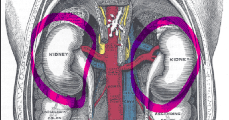
この書籍に寄稿しているマーティン・フリードリッヒという人物を調べていた所、腎炎の検査方法について論文を書いていることが分かった。
血清病による糸球体腎炎を追求している私からすると耳寄り情報があるかも…!と思って精読したら。尿検査の論文だった。
しかし、120年前の時点でこれだけのことが分かっていたのかと衝撃を受けたので、折角なので翻訳したものをそのまま記事にしたいと思う。
元リンクはコチラだ
CLEVELAND-Medical GAZETTE Vol.XVI.No.3.,Jan,1901
該当のページは198頁になる
THE LABORATORY DIAGNOSIS OF NEPHRITIS
腎炎の検査診断
The diagnosis of parenchymatous nephritis, be it acute or chronic, is made in the laboratory. The urinary changes are in the vast majority of cases pathognomonic. These changes affect the daily amount of the urine and its composition. The amount is less, the solids are relatively increased, raising the specific gravity and causing a turbidity by their insolubility in the scanty water. Abnormal ingredients appear, as renal epithelium, blood, albumin and casts.
柔組織腎炎の診断は、急性であれ慢性であれ、実験室で行われる。尿の変化は、ほとんどの場合、病的なものである。これらの変化は、1日の尿量とその組成に影響する。尿量は減少し、固形分が相対的に増加し、比重が増加し、乏しい水に溶解しないために濁りを生じる。腎臓上皮、血液、アルブミン、尿円柱などの異常な成分が現れる。
※尿円柱(urinary cast):リンク
遠位尿細管、集合管で形成される、Tamm-Horsfallムコ蛋白質とアルブミンが凝固沈殿したものを基質として、細胞あるいはその変性成分が封入された円柱状構造物。
→脂肪円柱:尿細管が脂肪変性したもの。ネフローゼ症候群、糖尿病性腎症
Epithelium of the uriniferous tubules points directly toward the kidneys. It is different with blood which may have an extra-renal origin. It is usually not difficult to distinguish between renal and extrarenal haematuria. In the former, the blood is intimately mixed with the urine, the red corpuscles are washed out, having lost almost all of the haemoglobin and presenting under the microscope a mere ring(shadow). In the latter, the erythrocytes are better preserved and clots are likely to form. The best means of discovering blood in the urine is the miroscope, the finding of red corpuscles eaves no doubt, while the spectroscope, chemical tests and Teichmann’s haemin crystals prove only the presence of the blood coloring matter which can get into the urine without hemorrhage as in cases of haemoglobinuria.
尿細管の上皮は直接腎臓のほうを向いている。腎臓以外に由来する可能性のある血液とは異なる。腎血尿と腎外血尿の区別は、通常、困難ではない。前者では、血液は尿と密接に混ざり合い、赤血球は洗い流され、ヘモグロビンのほとんどを失い、顕微鏡で見ると単なる輪(影)になっている。後者では、赤血球の保存状態がよく、血栓ができやすい。尿中の血液を発見する最良の方法は顕微鏡で、赤血球が見つかれば疑う余地はないが、分光器、化学検査、Teichmannのヘミン結晶は、ヘモグロビン尿の場合のように出血せずに尿中に混入しうる血液着色物質が存在することだけを証明するものである。
Albumin, like blood, can have a renal or entra-renal origin. The latter has its source in a pyclitis, eystitis or urethritis, or in the breaking of an absess into the urinary tract. The finding of puscorpuscles in the sediment, the absence of albumin in the filtered urine and the lack of urinary changes gives us the true diagnosis. Here need mentioning chyluria, which, be it parasitic or non-parasitic, is always accompanied, and haemoglobinuria which is usually preceded and followed by albuminuria.
アルブミンは血液と同じように、腎臓由来と腎臓内由来がある。後者は水晶体炎、膀胱炎、尿道炎、膿瘍の尿路破裂に起因する。尿沈渣に鱗屑を認め、濾過尿にアルブミンを認めず、尿路に変化を認めないことから、真の診断が可能である。寄生虫性、非寄生虫性にかかわらず常に伴うチル尿と、通常アルブミン尿の前後にみられるヘモグロビン尿に言及する必要がある。
Renal or true albuminuria was formerly considered as a sure symptom of Bright’s disease. Since the subject has been studied better, and especially since we have more delicate tests for the discovery of albumin, opinions have changed. Spiegler with his most delicate re-action which indicates albumin 1.35,000; Hg. Bichl. 8, Ac. Tart. 4, Aqu. Dest. 200, glycerine 20; states that it was difficult for him to find in the higher classes of society a sample of urine entirely free from albumin. Roch’s reagent, a saturated solution of sulpho-salicylic acid indicates albumin 1.50000. Such minimal quantities are of no clinical importance. It is called now physiological albuminuria. We find it after hard muscular exercise, heavy meals rich in albumen, cold baths, mental excitement and long continued brain work. Pavy’s cyclical albuminuria belongs to this category. This cycle is merely dependent upon external conditions. In the morning, after the night’s rest, albumin is less in any case of albuminuria and increases toward evening, after the days work. Besides the cycle can be broken by muscular exercise, cold baths, etc.
以前は、腎臓あるいは真のアルブミン尿は、ブライト病の確実な症状であると考えられていた。しかし、この問題がよりよく研究され、特にアルブミンを発見するためのより繊細なテストが行われるようになってから、意見が変わってきた。Spieglerは、アルブミン1.35,000を示す最も繊細な反応を示し、[Hg. Bichl. 8、Ac. Tart. 4、Aqu. Dest. 200、グリセリン20]社会の上流階級でアルブミンを全く含まない尿サンプルを見つけるのは困難であったと述べている。ロッシュの試薬、スルホサリチル酸の飽和溶液はアルブミンを1.50000と表示する。このような微量なアルブミンは臨床的には重要ではない。現在では生理的アルブミン尿と呼ばれている。激しい筋肉運動、卵白を多く含む重い食事、冷浴、精神的興奮、長時間にわたる頭脳労働の後に見られる。Pavyの周期性アルブミン尿はこの範疇に属する。この周期は、単に外的条件に左右されるだけである。朝、夜の休息後、アルブミンはどのようなアルブミン尿の場合でも少なくなり、夕方、日中の仕事の後に向かって増加する。さらに、このサイクルは、筋肉運動や冷浴などによって崩れることもある。
Virchow has mentioned already as far back as 1846, that the urine of new born during the first eight or ten days of life frequently shows albumin, which afterward disappears. Women in labor have usually traces of it.
ウィルヒョーは1846年の時点ですでに、生後8〜10日の新生児の尿にはしばしばアルブミンが含まれ、その後消失することを述べている。陣痛中の女性には通常その痕跡がある。
Nearer to a pathological condition but still without any detectable changes in the kidney, are the following cases of albuminuria:
病的な状態に近いが、まだ腎臓に検出可能な変化がない場合、次のようなアルブミン尿の症例がある。
1. Febrile albuminuria as in diphtheria, the eruptive fevers, typhus, typhoid, tetanus, cerebro-spinal meningitis, rheumatism, influenza.
2. Albuminuria in general non-febrile, morbid conditions in which the composition of the blood is affected as in anaemia(simple and pernicious) leukaemia, pseudo-leukaemia, scorbut, in some cases of icterus and diabetes melitus.
3.In affections of the nervous system as in epilepsy, delirium tremens, hysteria, cerebral hemorrhage, nervous exhaustion, migraine and Basedow’s disease.
1. ジフテリア、発疹熱、チフス、破傷風、脳脊髄膜炎、リウマチ、インフルエンザなどの発熱性アルブミン尿。
2. 貧血(単純および悪性)、白血病、偽白血病、壊血病、黄疸のいくつかのケースや黄糖尿病のように、血液の組成に影響を与える一般非熱病、病的状態におけるアルブミン尿。
3.てんかん、振戦せん妄、ヒステリー、脳出血、神経衰弱、片頭痛、バセドー病など神経系統の疾患。
Lastly albuminuria is found in certain affections of the intestines as in incarceration and in acute diarrhoeas.
最後に、アルブミン尿は、嵌頓や急性の下痢など、ある種の腸の病気で見られるものである。
嵌頓(かんとん:incarceration):腸や子宮などの内臓諸器官が、組織の隙間から脱出し、そのまま増大して腫れ上がり、もとにもどらなくなった状態
by コトバンク
The albumin of nephritic urine is the albumin of the blood, serum-albumin(serum) and serum-globullin(paraglobulin). We must remember that other albumins may find access to the urine as nucleo-albumin, the source of which are the nuclei of the cells. After febrile affections there is usually a great desquamation of renal epithelium and disintegration of nuclei from, which we may get a considerable amount of nucleo-albumin in the urine. The same is the case in a catarrhal condition of the bladder and a vesical nucleo-albuminuria has been described. Besides, we find albumose,hemi-albumose or propeptone which was first pointed out by Bence Jones in a case of osteomalacia and since in quite a few other diseases.
腎尿のアルブミンは、血液のアルブミン、血清アルブミン(血清)、血清グロブリン(パラグロブリン)である。その他のアルブミンは、細胞核を原料とするヌクレオアルブミンとして、尿中に混入することがあることを忘れてはならない。熱病の後では、通常、腎臓上皮の大きな剥離と核の崩壊があり、そこからかなりの量のヌクレオアルブミンが尿中に出てくることがある。膀胱のカタル性疾患でも同様で、小水疱性核酸アルブミン尿が報告されている。このほか、アルブモース、ヘミアルブモース、プロペトンなどがあるが、これはベンス・ジョーンズが骨軟化症の症例で最初に指摘し、その後、他の多くの疾患でも指摘されるようになった。
The tests commonly used for the detection of albumin in the urine are 1,heat; 2,nitric acid; 3,acetic acid plus ferrocyanide of potassium. All three have to be employed in every case in order to avoid errors. The old teaching, “get acquainted with one test and stick to it,” is faulty. Heat coagulates serum-albumin and paraglobulin. Other albumins as albumose, hemi-albumose and nucleo-albumin remain in solution. If we underlay urine with clear nitric acid, all the albumins present will coagulate, forming a white ring at the line of contact. In order to exclude albumose and hemi-albumose, we apply gentle heat. If the ring does not disappear, it can consist of serum and globulin or nucleo-albumin. The latter we exclude by using the third test, acetic acid plus ferrocyanide of potassium, a ten percent solution of each. On the addition of a drop or two of acetic acid nucleo-albumin coagulates in the form of a white cloud and can be filtered out. If now by adding a drop or two of a solution of ferrocyanide of potassium we find again a cloudiness, it must be serum-albumin and paraglobulin, the albumose and hemi-albumose having been excluded by application of heat to the nitric acid test.
尿中のアルブミンの検出には、1,熱、2,硝酸、3,酢酸とフェロシアン化カリウムがよく使われる検査方法である。この3つの検査は、間違いを避けるために、すべてのケースで使用されなければならない。昔から言われている「一つの検査に習熟し、それに固執する」というのは誤りである。熱は血清アルブミンとパラグロブリンを凝固させる。その他のアルブミン、ヘミアルブミン、ヌクレオアルブミンは溶液のままである。尿に透明な硝酸をかけると、アルブミンはすべて凝固し、接触した部分に白いリングを形成する。そこで、アルブミンとヘミアルブミンを除くために、穏やかに加熱する。リングが消えない場合は、血清とグロブリンまたはヌクレオアルブミンである可能性がある。後者は第3のテスト、酢酸とフェロシアン化カリウム、それぞれの10%溶液を用いて除外する。酢酸を1滴か2滴加えると、ヌクレオアルブミンは白い雲のような形で凝固し、濾過することができる。ここでフェロシアン化カリウムの溶液を1、2滴加えて再び白濁が見られたら、それは血清アルブミンとパラグロブリンに違いなく、アルブモースとヘミアルブモースは硝酸試験の熱で除かれたものである。
The most important factor after albumin are casts. They are subdivided into a.,blood casts formed by red blood corpuscles held together likely by the fibrin with here and there a leucocyte. They are found in parenchymatous nephritis, but also in renal hemorrhage.
アルブミンに次いで重要なのが尿円柱である。これらは次のように細分化される
a.赤血球がフィブリンによって結合され、白血球が混在する血液円柱である。柔組織腎炎に見られるが、腎出血にも見られる。
b. Cellular casts composed almost exclusively of renal epithelium which is in all stages of disintegration, from cloudy swelling to fatty degeneration. Their presence indicates that the disease affects the tubules and their absence as in post-scarlatinal nephritis, points toward a glommerular affection only.
b.白濁した腎臓腫大から脂肪性変性まであらゆる崩壊段階にある腎上皮のみからなる細胞性円柱。この細胞屑の存在は、この病気が尿細管に影響を及ぼしていることを示し、瘢痕化後腎炎のように細胞屑がない場合は、膠原病だけの病気であることを示唆する。
c. Granular casts, subdivided into coarse and fine granular, are now considered as cellular casts which have remained longer in the tubules. They are certainly, especially the fine granular, oftenest found in chronic parenchymatous nephritis.
c.顆粒円柱は粗粒と細粒に分けられ、現在では尿細管に長く留まっている細胞性円柱と考えられている。特に細粒型は、慢性柔組織腎炎で最もよく見られる。
d. Structureless casts, waxy and hyaline. We know little of the formation of the former. They are seldom found and give the reaction of amyloid, but have nothing to do with amyloid infiltration. The hyaline casts are the ones which caused formerly, so much trouble among the profession, as some considered them as formed by an exudate, therefore exudative nephritis. The prevailing opinion now is that they are derived from kidney epithelium. They appear in all forms of Bright’s disease, and once in a while are even found in apparently normal urine.
d.無構造円柱、ワックス状、ヒアルロン酸状。前者の形成についてはほとんど分かっていない。めったに見られず、アミロイドの反応を示すが、アミロイドの浸潤とは関係がない。ヒアルロン酸の円柱は、以前は滲出液によって形成される、つまり滲出性腎炎であると考える者がいて、専門家の間で大きな問題となったものである。現在では、腎臓の上皮に由来するというのが一般的な見解である。ブライト病のすべての病型に出現し、時には正常な尿の中にさえ見出される。
I have premised these statements concerning our present knowledge of albuminuria and casts in order to clear the ground for the diagnosis of parenchymatous nephritis.
私は、アルブミン尿と円柱に関する現在の知識を前提に、柔組織腎炎の診断の根拠を明らかにするために、このような記述をした。
The twenty-four hours’ urine must be conscientiously gathered. The amount is always below the normal, sometimes as low as 500 to 200cc. For a time we may have even complete anuria. The color is from a light red to a brownish dark hue. The urine is never clear. It is passed turbid and the sediment is heavy. The specific gravity is high 1022-1030. The urinary solids although relatively increased, are below the normal daily amount, as can easily be shown by Haeser’s co-efficient: 2.33. For chemical examination the urine must be filtered, and if very concentrated, it is highly advisable to dilute it one-half or two-thirds with distilled or at least filtered water, as then every test shows up better. The three tests will show a considerable amount of albumin. Esbach is likely to indicate ½ to 1 per cent. Esbach’s method is only approximative, but close enough for practical purpose.
24時間分の尿を斟酌する必要がある。その量は常に正常値より少なく、時には500ccから200ccにもなる。一時は完全な無尿になることもある。色は薄い赤から茶色がかった濃い色である。尿は決して透明ではない。濁った状態で通過し、沈殿物は重い。比重は1022-1030と高い。尿の固形分は比較的多いが、通常の1日量より少なく、ヘーザーの係数2.33で容易にわかる。化学的検査では、尿をろ過しなければならない。非常に濃縮されている場合は、蒸留水または少なくともろ過した水で2分の1から3分の1に希釈することを強くお勧めする。この3つの検査では、かなりの量のアルブミンが検出される。エスバッハは1/2から1パーセントを示すと思われる。エスバッハの方法は、あくまでも近似的なものだが、実用上は十分に近いものである。
Microscopy of the sediment demands a fresh sample of urine. The morning’s urine, except after a restless night with much tossing, shows the pathological elements the least, and the urine along in the day or towards evening is better, although any sample will usually do.
沈殿物の顕微鏡検査には、新鮮な尿のサンプルが必要である。朝の尿は、寝返りの多い夜の後を除けば、最も病的要素が少なく、日中または夕方の尿がよい。
Under the microscope we find usually red blood corpuscles, a few leucocytes, renal epithelium and casts, hyaline, epithelial and blood casts. In post-scarlatinal nephritis which affects commonly the glommeruli only, we find no epithelium nor epithelial casts.
顕微鏡で見ると、通常、赤血球、少数の白血球、腎臓上皮と尿円柱、ヒアルロン酸、上皮、血液円柱があることがわかる。扁平上皮後腎炎では、上皮も上皮性円柱も認められない。
All the morbid changes of the urine taken together, viz., turbidity, less amount with higher specific gravity than normal, the presence of a rather high per cent. of albumin with red blood corpuscles, epithelial cells and casts in the sediment, are pathognomonic of parenchymatous nephritis, and we can safely say that it is an acute attack. Where the specific gravity sinks closer down to the normal or even becomes normal and the twenty-four hours’ amount approaches nearer the amount passed in health during the same time with a scarcity of epithelial but a great many granular, especially fine granular casts, we have a right to think that the process is chronic. But the two stages cannot be separated by sharp lines, neither by clinician nor microscopist.
尿の病的な変化、すなわち濁り、量が少なく比重が大きいこと、アルブミンが赤血球、上皮細胞、沈殿物中にかなり高い割合で存在することは、柔組織腎炎の病徴であり、急性発作であると言って差し支えない。比重が正常値に近づき、あるいは正常値になり、24時間の量が健康時の量に近づき、上皮細胞は少ないが粒状、特に細かい粒状の円柱が多い場合は、慢性の経過と考えるのが妥当であろう。しかし、この2つの段階は、臨床医も顕微鏡医も、はっきりとした線で分けることはできない。
Of other morbid conditions renal hemorrhage resembles acute nephritis the closest. In both we have albumin, blood and blood-casts, but in hemorrhage the amount of urine is normal, and in the sediment we find no renal epithelia nor hyaline and epithelial casts. One day’s urine may be full of blood, the next day’s sample entirely free from it. In chronic congested kidney(Stauungsniere) we get less urine than normal, with a higher specific gravity and a small amount of albumin, but no blood, no renal epithelia nor casts. An amyloid kidney gives albuminous urine, but the amount is higher than normal, the specific gravity lower, and the urine is clear, forming little or no sediment. In interstitial nephritis we have also a great deal more urine than normal with a low specific gravity. Albumin may or may not be present, so that we cannot make the diagnois of contracted kidney(Schrumpfniere) from the examination of the urine alone. We have to take into account the cardio-vascular changes.
他の病的状態の中で、腎出血は急性腎炎に最もよく似ている。両者ともアルブミン、血液、血液円柱を認めるが、腎出血では尿量は正常であり、沈殿物には腎上皮もヒアルロン酸や上皮円柱も認められない。ある日の尿に血液が多くても、翌日の検体には全く血液がないこともある。慢性うっ血性腎臓(Stauungsniere)では、通常より尿量が少なく、比重が大きく、少量のアルブミンを含むが、血液、腎上皮、尿円柱は認められない。アミロイド腎では、アルブミン尿が出るが、量は通常より多く、比重は小さく、尿は透明で、沈殿物はほとんどできない。間質性腎炎でも、通常より多量の尿が出るが、比重は小さくなる。アルブミンの有無は関係せず、尿の検査だけでは縮瞳腎と診断することはできない。心・血管系の変化も考慮しなければならない。
サポートで生き長らえます。。。!!